Sudden Cardiac Arrest vs Heart Attack
April 10, 2024
What is the Indication for Mouth-To-Mouth Rescue Breaths?
April 29, 2024Table of contents
- What is Cardiac MRI?
- How Does Cardiac MRI Work?
- Difference Between a Cardiac MRI and an MRI
- When is a Cardiac MRI Performed?
- What Does a Cardiac MRI Diagnose?
- Benefits of Cardiac MRI
- Limitations & Risks of Cardiac MRI
- Preparing for a Cardiac MRI Exam
- What to Expect During a Cardiac MRI Exam?
- Why Choose Cardiac MRI?
- People Also Asked About Cardiac MRI
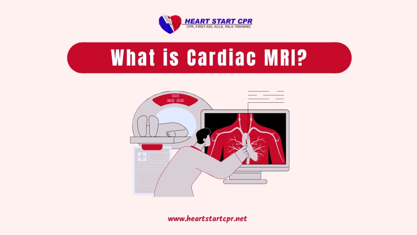
What is Cardiac MRI?
Cardiac MRI or Cardiovascular Magnetic Resonance imaging (CMR) is a non-invasive medical imaging test that uses powerful magnetic fields, radio waves, and advanced computer technology to visualize the anatomy and functional information of the heart without surgery. It can help identify issues such as heart muscle damage from a heart attack, structural problems with the heart’s chambers, diseases of the large blood vessels (such as the aorta), and congenital heart defects in both adults and children. It is particularly useful for its ability to produce high-resolution, three-dimensional images that can be viewed from multiple angles, offering a comprehensive evaluation of cardiac health.
How Does Cardiac MRI Work?
Cardiac MRI works on the basic principle of nuclear magnetic resonance. When a patient is placed inside a powerful magnetic field, the hydrogen atoms in the body’s tissues get aligned. Radiofrequency pulses are then applied which excite these aligned hydrogen atoms. As the excited hydrogen atoms return to their normal unexcited state, they release energy in the form of radio waves.
These emitted radio waves are unique for each type of tissue in the body. A receiver coil detects these signals which are then processed by a computer to construct detailed images of the heart and surrounding structures. By acquiring images in different planes (axial, coronal and sagittal) and during different phases of the cardiac cycle, cardiologists can assess the anatomy and function of the heart with great accuracy.
Difference Between a Cardiac MRI and an MRI
Here is a table comparing the main differences between a Cardiac MRI and a regular MRI:
| Basis | Cardiac MRI | MRI |
| Purpose | To image the heart and blood vessels for signs of heart diseases like cardiomyopathy, heart defects, damages etc. | To image various parts of the body like brain, joints, abdomen etc. for signs of diseases or injuries. |
| Focus | Specifically focuses on heart function, anatomy and blood flow within the heart and surrounding vessels | Depends on the area being imaged but it is not specifically focused on the heart. |
| Technique | Uses specialized techniques like ECG gating and fast imaging sequences to “freeze” cardiac motion and blood flow | Standard MRI techniques without ECG gating as motion is not an issue |
| Scan time | Longer scan time (30-90 mins) as multiple sequences required to view heart from different angles | Shorter scan time (10-30 mins) depending on area scanned |
| Image quality | Provides high resolution images of the heart due to specialized coils and techniques | Image quality depends on area scanned but may not be as detailed as cardiac MRI for heart imaging |
| Positioning | Patient is positioned feet-first and head-first inside the bore. | Positioning depends on the body part to be imaged but usually head or feet first. |
| Interpreting Doctor | Specialized cardiac radiologists and cardiologists. | Radiologists with training in MRI interpretation. |
When is a Cardiac MRI Performed?
Your doctor can order a cardiac MRI for several reasons related to assessing, diagnosing, or monitoring heart disease. Some common uses include:
1. Evaluating Heart Function
MRI provides precise measurements of the heart’s pumping ability, blood flow, and overall performance that are vital for detecting issues like:
- Heart failure
- Coronary artery disease
- Heart valve disorders
- Structural abnormalities
- Cardiac shunts
- Cardiac tumors
2. Assessing Heart Anatomy
The high-resolution anatomical images from MRI help visualize and pinpoint problems with the heart’s physical structure, such as:
- Myocardial viability after heart attack
- Heart muscle inflammation or scarring
- Cardiac chamber enlargement
- Heart wall thickening or thinning
- Congenital heart defects
- Pericardial diseases
3. Guiding Treatment Planning
Cardiac MRI gives doctors detailed visuals to plan treatments and procedures by clearly revealing:
- Extent and location of damage or blockages
- Valve abnormalities requiring repair/replacement
- Areas to target interventions or cardiac surgery
- Pre-transplant evaluation of heart status
4. Monitoring Conditions Over Time
With its non-invasive nature, MRI can be performed repeatedly to safely track changes and disease progression, such as:
- Coronary artery disease progression
- Effectiveness of medical/interventional treatments
- Post-operative recovery after heart procedures
- Heart transplant monitoring for complications
What Does a Cardiac MRI Diagnose?
Cardiac MRI provides invaluable diagnostic information about a wide range of heart conditions, including:
1. Heart Function
- Ejection fraction (pumping ability)
- Wall motion abnormalities
- Valve disorders impacting flow
- Congenital shunts and malformations
- Heart failure severity
2. Coronary Artery Disease
- Reduced blood flow to the heart
- Locations of arterial blockages
- Scarring from prior heart attacks
- Viability of heart muscle tissue
3. Structural/Anatomical Issues
- Chamber sizes and wall thickness
- Muscle inflammation or scarring
- Structural defects like holes/malformations
- Heart muscle masses or tumors
- Pericardial diseases like fluid buildup
Benefits of Cardiac MRI
As an advanced cardiac imaging modality, MRI offers several benefits compared to other tests:
- No ionizing radiation exposure (safer than x-rays/CT)
- Unmatched soft tissue contrast and resolution
- Can capture dynamic 3D visuals of heart function/mechanics
- Precise evaluation of myocardial viability after heart attack
- Detects acute and chronic coronary artery disease
- Identifies heart muscle abnormalities like inflammation
- Helpful for evaluating complex congenital heart disease
- Can image from any orientation without obstruction
- Non-invasive and generally well-tolerated procedure
Limitations & Risks of Cardiac MRI
While a powerful diagnostic tool, cardiac MRI is not appropriate or necessary for all patients and heart-related issues. Some key limitations include:
- Not suitable for patients with non-compatible implanted devices
- Can be challenging for patients with claustrophobia or obesity
- Longer scan times required (up to 1 hour) compared to other tests
- Higher costs compared to basic tests like echocardiograms
- Unable to capture live x-ray/fluoroscopic-like images
- Sometimes lower resolution images in patients who can’t breath-hold
There are also some potential risks to be aware of with MRI exams, although they are relatively rare:
- Allergic reaction to contrast material (gadolinium) if used
- Nerve stimulation from rapidly pulsating magnetic fields
- Displacement of implanted device if not MRI-compatible
Overall, cardiac MRI is considered a very safe procedure when properly screened and monitored by trained technicians and radiologists.
Preparing for a Cardiac MRI Exam
Proper preparation is required before the test to ensure diagnostic-quality images are obtained and the scanning process goes smoothly. Here are some common guidelines:
- You may be asked to avoid eating for 2-4 hours before the exam to prevent artifacts from digestive gases. Drink only clear fluids.
- Inform technicians about any medical devices or implants (pacemakers, defibrillators, etc.) which are contraindicated for MRI.
- Remove all metallic objects, such as watches, jewelry, eyeglasses, etc., as they can interfere with the strong magnetic field.
- Loose-fitting, comfortable clothes without metal zippers should be worn. You may be given a hospital gown.
- Some patients need an IV line placed to inject a contrast agent (gadolinium) to enhance image quality.
- Blood work may be ordered to check kidney function before contrast is used, as it is eliminated by the kidneys.
- You should plan to remain very still during imaging, which takes 30-90 minutes. Bring along music or games on your phone.
What to Expect During a Cardiac MRI Exam?
On the day of the test, here is generally what happens during a typical Cardiac MRI study:
- You will be asked to change into a hospital gown, and all metal objects will be removed.
- EKG electrodes are attached to monitor your heart rhythm continuously during scanning.
- Lying on the MRI table, safety screening for pacemakers, etc., is done using a wand before entering the scanner room.
- Earplugs and headphones are provided to minimize noise from the loud knocking MRI sounds. You may be given a call button.
- Breath-holding instructions will be explained – you need to stay motionless for 10-20 seconds during acquisitions.
- The table glides into the open-ended cylindrical bore of the scanner, which is about 1.5 meters long.
- Series of scans will then be performed as you lie still. Contrast may be administered during this phase via the IV.
- Simple instructions like “breathe” or “hold breath” will be given via intercom during the 30-90 minute exam.
- Images are swiftly generated on the computer console in the control room and examined by the interpreting physician.
- You will then be assisted out of the machine. Results are usually available in 1-3 days.
Why Choose Cardiac MRI?
Cardiac MRI is a powerful non-invasive imaging technology that provides detailed anatomical and functional information about the heart and vasculature without using any ionizing radiation. Its versatility, soft tissue contrast, and physiological evaluation capabilities have made it a valuable diagnostic and clinical management tool for diverse cardiac disorders. By providing comprehensive information, cardiac MRI aids accurate diagnosis, treatment planning and assessment of disease progression – ultimately helping improve quality of cardiovascular care.
People Also Asked About Cardiac MRI
Is cardiac MRI safe?
Yes, cardiac MRI is a highly safe imaging technique when used correctly and on selected patients. The magnetic and radiofrequency fields do not cause any biological harm. However, patients with pacemakers, certain implants or metal foreign bodies cannot undergo MRI due to safety concerns.
Is there any radiation exposure with MRI?
No, MRI does not use any ionizing radiation like X-rays, CT scans, or nuclear medicine procedures. The images are formed entirely by manipulating radiofrequency and strong magnetic fields with no radiation exposure involved.
How long does a Cardiac MRI take?
A standard MRI of the heart usually takes half an hour to an hour and a half from entering the scanner to completion, depending on the number of images/sequences needed. Emergency/urgent exams can sometimes be completed faster.
Is Cardiac MRI painful?
Cardiac MRI is painless. You can experience some discomfort from having to remain still inside the tight scanner bore or due to anxiety, but no pain is caused by the actual imaging process.
Are results conclusive?
When performed and interpreted by experienced staff, cardiac MRI findings are very reliable for diagnosis and tracking disease. However, in some situations other supporting tests may still be needed for complete evaluation or management decisions.

